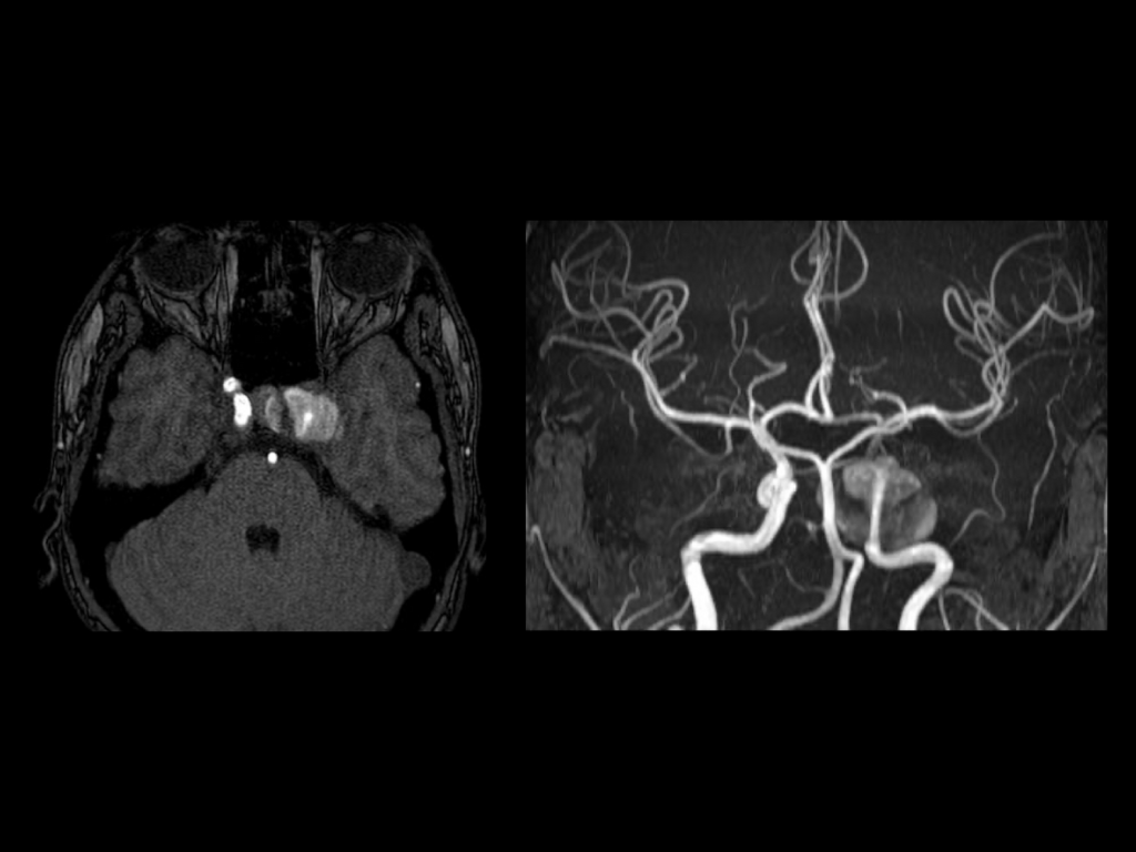
Axial, sagittal, coronal T2, axial T1, MRA images reveal a large 2.8 x 2.3 cm outpouching showing flow related enhancement arising from cavernous segment of left internal carotid artery. MRI reveals mixed signal intensities representing various stages of clot in the thrombosed portion of the lumen; evidence of flow within the patent portion of the residual lumen; periluminal high signal around the patent residual lumen, reflecting either slow flow or methemoglobin.
Diagnosis
Giant partially thrombosed ICA aneurysm
Summary
Giant intracranial aneurysms are defined as those with diameters of 25 mm or more and represent about 5% of all intracranial aneurysms. Due to its size, a giant aneurysm is responsible for intracranial mass effect rather than intracranial hemorrhage as seen in the index case. The treatment may be either surgical, endovascular or both.
Further reads
Borni, M., Kolsi, F., Cherif, I., & Boudawara, M. Z. (2022). A giant partial thrombosed aneurysm of the internal cavernous carotid artery mimicking a meningioma of the lesser wing of the sphenoid bone. Radiology case reports, 17(4), 1325–1329. https://doi.org/10.1016/j.radcr.2022.01.075